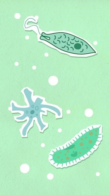
Cellular transport‚ encompassing diffusion and active mechanisms‚ is fundamental for life‚ moving materials in and out of cells as detailed in recent studies.
Importance of Cellular Transport
Cellular transport is absolutely vital for all life forms‚ ensuring cells receive essential nutrients while expelling waste products. This dynamic process maintains cellular homeostasis‚ crucial for proper function and survival. Without effective transport‚ cells cannot perform necessary metabolic reactions‚ synthesize proteins‚ or respond to environmental cues.
The movement of molecules‚ whether through passive or active mechanisms‚ directly impacts cellular signaling‚ nutrient uptake‚ and waste removal. Researchers are increasingly focused on understanding these intricate processes‚ particularly peptide transport mechanisms and the role of membrane proteins.
Recent advancements‚ like utilizing DNA origami to control membrane function‚ highlight the growing sophistication in studying and manipulating cellular transport. Understanding these mechanisms is paramount for addressing various biological and medical challenges‚ from developing targeted drug delivery systems to comprehending disease pathogenesis.
Cell Membrane Structure and its Role in Transport
The cell membrane‚ a phospholipid bilayer with embedded proteins‚ acts as a selective barrier controlling the passage of substances in and out of the cell. This structure dictates how molecules utilize mechanisms like diffusion‚ facilitated diffusion‚ and active transport to cross. The membrane’s fluidity‚ influenced by cholesterol and protein content‚ impacts transport efficiency.
Membrane proteins‚ including channels and carriers‚ are integral to facilitated diffusion and active transport‚ providing pathways for specific molecules. Recent research demonstrates that manipulating membrane structure‚ even with techniques like DNA origami‚ can directly influence transport rates and selectivity.
The membrane’s structure isn’t static; it dynamically adjusts to maintain cellular homeostasis. Understanding this interplay between structure and function is crucial for comprehending how cells manage nutrient uptake and waste removal‚ processes vital for survival.

Passive Transport Mechanisms
Passive transport‚ including simple diffusion and facilitated diffusion‚ moves substances across membranes without energy expenditure‚ relying on concentration gradients.
Simple Diffusion
Simple diffusion represents the most basic mechanism for traversing the plasma membrane‚ requiring no assistance from membrane proteins. Molecules simply dissolve into the lipid bilayer and move from areas of high concentration to areas of low concentration‚ following a concentration gradient.
This passive process continues until equilibrium is reached‚ ensuring equal distribution. The rate of diffusion is influenced by factors like temperature‚ molecule size‚ and the membrane’s permeability. Small‚ nonpolar molecules‚ such as oxygen and carbon dioxide‚ readily diffuse across the membrane.
However‚ polar and charged molecules face significant barriers due to the hydrophobic core of the lipid bilayer‚ limiting their ability to utilize this straightforward transport method. It’s a fundamental process for essential gas exchange within organisms.
Facilitated Diffusion

Facilitated diffusion alleviates the limitations of simple diffusion for polar and charged molecules‚ utilizing membrane proteins to assist their passage. This passive transport method still relies on a concentration gradient‚ meaning no cellular energy expenditure is required. However‚ it necessitates the aid of specialized proteins to overcome the hydrophobic barrier of the lipid bilayer.
Two primary types of proteins mediate facilitated diffusion: channel proteins and carrier proteins. Both enhance the membrane’s permeability to specific solutes‚ enabling their efficient transport. This process is crucial for the uptake of essential nutrients and the removal of waste products‚ ensuring cellular homeostasis.
The specificity of these proteins ensures that only appropriate molecules are transported‚ maintaining cellular integrity and function.
Channel Proteins
Channel proteins form hydrophilic pores across the cell membrane‚ allowing specific ions or small polar molecules to pass through rapidly. These proteins don’t bind to the transported solute; instead‚ they provide a continuous pathway‚ accelerating diffusion down the concentration gradient.
Channel proteins exhibit selectivity based on size and charge‚ ensuring only appropriate molecules traverse the membrane. Some channels are ‘gated‚’ opening or closing in response to specific signals like voltage changes or ligand binding‚ regulating transport based on cellular needs.
Aquaporins‚ for example‚ are channel proteins facilitating rapid water transport. Their existence dramatically increases membrane permeability to water‚ vital for maintaining cell turgor and fluid balance.
Carrier Proteins
Carrier proteins bind to specific solutes and undergo conformational changes to transport them across the cell membrane. Unlike channel proteins‚ carriers exhibit specificity and saturation kinetics – meaning they can only bind certain molecules and have a limited number of binding sites.
This binding process initiates a shape alteration in the protein‚ effectively shuttling the solute to the other side of the membrane‚ where it’s released. Carrier proteins facilitate both facilitated diffusion (passive) and active transport‚ depending on the energy requirements.
The rate of transport is influenced by the concentration of both the solute and the carrier protein‚ showcasing a distinct transport mechanism compared to simple diffusion or channel-mediated transport.

Active Transport Mechanisms
Active transport requires energy to move substances against their concentration gradients‚ utilizing primary and secondary mechanisms for cellular material movement.
Primary Active Transport
Primary active transport directly utilizes metabolic energy‚ typically in the form of ATP hydrolysis‚ to drive the movement of molecules across cell membranes. This process establishes and maintains electrochemical gradients essential for various cellular functions. Unlike secondary active transport‚ it doesn’t rely on the transport of another molecule.
A prime example is the sodium-potassium pump‚ a crucial mechanism found in animal cells. This pump expends ATP to move sodium ions out of the cell and potassium ions into the cell‚ against their respective concentration gradients. This action is vital for maintaining cell volume‚ nerve impulse transmission‚ and secondary active transport processes.
Furthermore‚ proton pumps are another significant type of primary active transport‚ commonly found in plant and fungal cells‚ contributing to nutrient uptake and pH regulation. These pumps actively transport protons (H+) across membranes‚ creating an electrochemical gradient used for driving other transport processes.
Secondary Active Transport
Secondary active transport leverages the electrochemical gradient established by primary active transport to move molecules across the cell membrane. It doesn’t directly consume ATP; instead‚ it utilizes the potential energy stored in the gradient created by pumps like the sodium-potassium pump. This transport relies on the simultaneous movement of two substances.
There are two main types: symport and antiport. Symport involves the movement of both substances in the same direction‚ while antiport moves them in opposite directions. For instance‚ sodium-glucose cotransporter utilizes the sodium gradient to transport glucose into the cell‚ even against its concentration gradient.
This mechanism is crucial for nutrient absorption in the intestines and kidneys‚ as well as for maintaining cellular homeostasis. It’s a highly efficient way to transport molecules‚ capitalizing on the energy already invested by primary active transport systems‚ showcasing the interconnectedness of cellular processes.
Sodium-Potassium Pump
The sodium-potassium pump (Na+/K+ ATPase) is a quintessential example of primary active transport‚ critically maintaining cellular resting potential and regulating cell volume. This integral membrane protein utilizes ATP hydrolysis to transport three sodium ions (Na+) out of the cell and two potassium ions (K+) into the cell‚ against their respective concentration gradients.
This process is vital for nerve impulse transmission‚ muscle contraction‚ and maintaining osmotic balance. The pump’s activity generates an electrochemical gradient‚ essential for secondary active transport and numerous cellular functions. It’s ubiquitously found in animal cells‚ demonstrating its fundamental importance.
Inhibition of the sodium-potassium pump can have severe consequences‚ disrupting cellular homeostasis and leading to various physiological dysfunctions‚ highlighting its crucial role in maintaining life.

Transport of Large Molecules
Endocytosis and exocytosis are crucial processes for moving large molecules‚ particles‚ or even cells across the cell membrane‚ facilitating essential cellular functions.
Endocytosis
Endocytosis represents a vital cellular process where substances are brought into the cell by enveloping them within the cell membrane‚ forming vesicles. This inward budding process allows cells to internalize molecules too large for direct diffusion or active transport mechanisms. Two primary forms of endocytosis exist: phagocytosis and pinocytosis‚ each serving distinct purposes.
Essentially‚ the cell membrane invaginates‚ surrounding the target material‚ and then pinches off to create an intracellular vesicle containing the engulfed substance. This vesicle then travels within the cell‚ delivering its contents to appropriate destinations for processing or storage. Endocytosis is critical for nutrient uptake‚ immune defense‚ and cellular communication‚ demonstrating its broad importance in biological systems.
Phagocytosis

Phagocytosis‚ often termed “cell eating‚” is a specialized form of endocytosis primarily utilized by immune cells like macrophages and neutrophils. This process involves the engulfment of large particles‚ such as bacteria‚ cellular debris‚ or foreign substances‚ exceeding one micrometer in diameter. The cell membrane dramatically extends pseudopodia – temporary cytoplasmic projections – to surround and internalize the target particle.
Once enclosed within a vesicle called a phagosome‚ the particle is then fused with a lysosome‚ an organelle containing digestive enzymes. These enzymes break down the engulfed material‚ effectively eliminating pathogens or clearing cellular waste. Phagocytosis is a crucial component of the innate immune response‚ defending the body against infection and maintaining tissue homeostasis.
Pinocytosis
Pinocytosis‚ meaning “cell drinking‚” is another type of endocytosis‚ but unlike phagocytosis‚ it involves the uptake of extracellular fluid containing dissolved solutes and small molecules. The cell membrane invaginates‚ forming small vesicles that pinch off into the cytoplasm‚ bringing the fluid and its contents inside. This process isn’t specific; cells essentially sample the surrounding environment‚ taking in whatever is present in the extracellular fluid.
Pinocytosis is a continuous process essential for various cellular functions‚ including nutrient acquisition and maintaining cellular hydration. It’s a non-selective process‚ meaning the cell doesn’t discriminate between different solutes. This contrasts sharply with receptor-mediated endocytosis‚ a more targeted form of pinocytosis.
Exocytosis
Exocytosis is the process by which cells transport large molecules‚ such as proteins and polysaccharides‚ out of the cell. Vesicles containing these substances fuse with the plasma membrane‚ releasing their contents into the extracellular space. This process is crucial for secretion‚ delivering signaling molecules‚ and expelling waste products.
There are two main types of exocytosis: constitutive and regulated. Constitutive exocytosis occurs continuously‚ while regulated exocytosis requires a specific signal‚ like a hormone or neurotransmitter. Both types involve vesicle trafficking and membrane fusion. Exocytosis is vital for cellular communication‚ growth‚ and maintaining the extracellular matrix.

Role of Membrane Proteins in Transport
Membrane proteins‚ including channels and carriers‚ are essential for peptide transport and controlling membrane function‚ as demonstrated by DNA origami research.
Peptide Transport Mechanisms

Peptide transport across cell membranes is a crucial process‚ heavily researched for its role in nutrient uptake and intracellular signaling. Understanding these mechanisms is vital‚ as peptides aren’t easily transported due to their charge and polarity.
Specific membrane proteins facilitate this transport‚ employing various strategies. Some peptides utilize carrier proteins‚ undergoing conformational changes to shuttle them across the lipid bilayer. Others might leverage channel proteins‚ forming hydrophilic pores for passage.
Recent advancements‚ like those utilizing DNA origami‚ offer potential for controlling and studying these transport processes with greater precision. Researchers are actively investigating how manipulating membrane structure impacts peptide translocation efficiency‚ potentially leading to novel therapeutic strategies and improved drug delivery systems.
Influence of DNA Origami on Membrane Function
DNA origami‚ a nanotechnology technique‚ is emerging as a powerful tool to precisely control the structure and function of biological membranes. Scientists at the University of Stuttgart have demonstrated the ability to manipulate membrane organization using this method‚ offering unprecedented control over cellular processes.
By designing specific DNA structures that interact with membrane components‚ researchers can create artificial membrane domains‚ influencing protein localization and transport efficiency. This approach allows for the investigation of how membrane architecture impacts various cellular functions‚ including peptide transport and signaling pathways.
The potential applications are vast‚ ranging from creating artificial cells with tailored properties to developing novel drug delivery systems and biosensors. DNA origami provides a versatile platform for studying and engineering membrane behavior at the nanoscale.

The Role of Phosphate in Cellular Transport
Phosphate (Pi) plays a crucial and multifaceted role in cellular transport processes‚ extending beyond its well-known function in energy metabolism. It’s essential for bioenergetics‚ powering transport via ATP and GTP‚ and regulates metabolic pathways like glycolysis and oxidative phosphorylation‚ directly impacting ion and molecule movement.
Phosphate groups are frequently involved in the phosphorylation of membrane proteins‚ altering their conformation and activity‚ thereby modulating transport rates. This post-translational modification is a key regulatory mechanism for numerous transporters.
Furthermore‚ phosphate itself is actively transported across cell membranes‚ often coupled to other ion transport‚ maintaining cellular phosphate homeostasis. Understanding phosphate’s influence is vital for comprehending overall cellular transport efficiency and regulation.