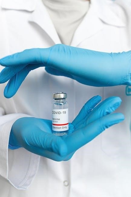
Sever’s disease, a common heel pain in active adolescents, is detailed in freely available guides like downloadable PDFs․
These resources outline causes, symptoms, and comprehensive treatment approaches for this condition․
What is Sever’s Disease?
Sever’s disease, or calcaneal apophysitis, isn’t a true disease but an inflammation of the growth plate in the heel․ This growth plate is a vulnerable area where tendons attach to the developing bone․ Frequently detailed in accessible PDFs, it primarily affects active children and adolescents during growth spurts․ Repetitive stress, particularly from sports involving running and jumping, contributes to micro-injuries․
These injuries cause heel pain, and understanding the condition is the first step towards effective management, as outlined in many treatment guides․
Prevalence and Demographics
Sever’s disease predominantly affects physically active adolescents, typically between the ages of 8 and 14, coinciding with rapid growth․ While precise prevalence rates vary, it’s considered one of the most common causes of heel pain in this age group․ Information readily available in treatment PDFs indicates a slightly higher incidence in boys, potentially due to their participation in more high-impact sports․
However, both genders are susceptible, and any child involved in repetitive running or jumping activities is at risk․
Historical Context & Naming
Sever’s disease is named after James Sever, an American orthopedic surgeon who first described the condition in 1912․ His initial observations detailed cases of heel pain in growing children, linking it to traction apophysitis of the calcaneal tubercle – the growth plate in the heel bone․
Treatment PDFs often acknowledge this historical context, noting that while the understanding of the pathophysiology has evolved, Sever’s original description remains foundational․
Understanding the Pathophysiology
Sever’s disease arises from repetitive stress on the immature calcaneal apophysis, a growth plate vulnerable to tension during activity, as detailed in treatment PDFs․
Growth Plate Involvement
Sever’s disease specifically impacts the calcaneal apophysis, a growth plate located at the back of the heel․ This area is a cartilaginous region where bone growth occurs in children and adolescents․ Treatment PDFs emphasize that repetitive stress, particularly from running and jumping, causes microtrauma to this vulnerable site․
Unlike fractures, it’s not a break in the bone itself, but inflammation where the Achilles tendon inserts․ Because the growth plate is weaker than mature bone, it’s susceptible to irritation and pain during periods of rapid growth and increased physical activity․ Understanding this growth plate involvement is crucial for effective treatment strategies, as outlined in available resources․
Biomechanical Factors & Stress
Treatment PDFs highlight how biomechanical factors significantly contribute to Sever’s disease․ Pronation, or the inward rolling of the foot, increases stress on the heel․ Inadequate footwear and training surfaces exacerbate this stress․ Repetitive impact during activities like running and jumping places considerable strain on the calcaneal apophysis․
Muscle imbalances, particularly tight calf muscles, also play a role by increasing tension on the Achilles tendon and, consequently, the heel․ Addressing these biomechanical issues through appropriate footwear, orthotics, and stretching exercises is a cornerstone of treatment, as detailed in comprehensive guides․
Role of Repetitive Impact
Treatment PDFs consistently emphasize the crucial role of repetitive impact in the development of Sever’s disease․ Activities involving frequent running, jumping, and quick changes in direction – common in sports – subject the growth plate to microtrauma․ This repeated stress overwhelms the apophysis’s capacity for repair, leading to inflammation and pain․
Without adequate rest and recovery, this cycle continues, worsening symptoms․ Guides often recommend activity modification to reduce impact, alongside other interventions․ Understanding this impact-related pathology is vital for effective management and preventing chronic issues․
Symptoms of Sever’s Disease
Treatment PDFs detail heel pain, especially during and after activity, as a primary symptom․ Tenderness and swelling around the heel are also frequently reported indicators․
Common Pain Locations
Treatment PDFs consistently pinpoint the posterior heel as the primary location for pain in Sever’s disease․ This discomfort arises from the growth plate at the back of the heel bone (calcaneus)․ Pain is often experienced directly beneath the heel, and can radiate outwards․
Adolescents frequently report pain when the heel is squeezed from the sides․ The pain isn’t typically on the sole of the foot, but rather focused on the back portion, where the Achilles tendon inserts․ Detailed guides emphasize localized tenderness as a key diagnostic feature, differentiating it from other foot ailments․
Pain Characteristics (Intensity, Timing)
Treatment PDFs describe the pain as typically worsening with activity, particularly running and jumping․ It’s often gradual in onset, not a sudden injury․ Intensity varies; some experience mild discomfort, while others report sharp, debilitating pain․
Pain is frequently most noticeable after activity, and may lessen with rest․ Morning pain is common, decreasing as the day progresses with gentle movement, only to return with increased physical exertion․ Guides highlight that pain levels fluctuate, and aren’t constant, making diagnosis challenging․
Associated Symptoms (Swelling, Tenderness)
Treatment PDFs consistently mention localized tenderness to the touch directly on the heel bone, specifically where the Achilles tendon inserts․ Mild swelling may be present around the heel, though it’s not always prominent․ Some individuals report tightness in the calf muscles, contributing to the biomechanical stress․
Importantly, PDFs emphasize the absence of fever or other systemic symptoms, differentiating Sever’s from infection․ Limping may occur, particularly after activity, as a protective mechanism to avoid exacerbating the heel pain․
Diagnosis of Sever’s Disease
Treatment PDFs highlight diagnosis via physical exam, focusing on heel palpation and assessing calf flexibility․ Imaging, like X-rays, is usually unnecessary unless other issues are suspected․
Physical Examination Techniques
Treatment PDFs emphasize a thorough physical exam is central to diagnosing Sever’s disease․ Palpation of the posterior calcaneus (heel bone) is key, eliciting pain is a strong indicator․ Assessing the child’s gait and range of motion in the ankle and foot is crucial․
Clinicians will evaluate calf muscle tightness, as this contributes to biomechanical stress․ Neurological examination helps rule out other potential causes of heel pain․ Documenting the location and intensity of pain during specific activities aids in accurate diagnosis, as detailed in available resources․
Ruling Out Other Conditions (Stress Fracture, Tendonitis)
Treatment PDFs highlight the importance of differential diagnosis․ Stress fractures present with more localized, persistent pain, often worsening with rest․ Tendonitis, like Achilles tendonitis, typically causes pain higher up the leg, not directly on the heel growth plate․
Careful questioning about the onset and nature of pain helps distinguish Sever’s from these conditions․ A physical exam assessing for point tenderness and swelling is vital․ If suspicion remains, imaging studies, as outlined in guides, may be necessary for definitive exclusion․
Imaging Considerations (X-rays, MRI ⎼ when necessary)
Treatment PDFs generally indicate that imaging isn’t routinely needed for Sever’s disease diagnosis․ X-rays may be used to rule out other issues like stress fractures, showing the growth plate’s appearance but not inflammation․
MRI scans are rarely necessary, reserved for cases with atypical presentations or diagnostic uncertainty․ They can visualize soft tissues, confirming the absence of tendon or ligament damage․ Guides emphasize clinical assessment as primary, with imaging as a secondary, selective tool․
Non-Surgical Treatment Options
Treatment PDFs consistently highlight conservative approaches: rest, ice, compression, and over-the-counter pain relievers are foundational for managing Sever’s disease symptoms effectively․
Rest and Activity Modification
Treatment PDFs emphasize that initial management of Sever’s disease centers on reducing activities that aggravate heel pain․ This doesn’t necessitate complete immobilization, but a temporary decrease in high-impact exercises like running and jumping is crucial․
Activity modification involves identifying and avoiding movements that exacerbate symptoms, potentially substituting them with low-impact alternatives such as swimming or cycling․
Gradual resumption of activities, guided by pain levels, is recommended, preventing re-injury and promoting healing․ Consistent rest periods during and after activity are also vital components of this approach․
Ice and Compression
Treatment PDFs consistently recommend applying ice packs to the affected heel for 15-20 minutes several times daily, particularly after activity․ This helps reduce inflammation and alleviate pain associated with Sever’s disease․
Compression, utilizing an elastic bandage, can further minimize swelling and provide support to the heel․ It’s crucial to avoid wrapping too tightly, ensuring continued circulation․
Combining ice and compression offers synergistic benefits, effectively managing acute symptoms and promoting a faster recovery process, as detailed in numerous downloadable guides․
Pain Management (Over-the-Counter Medications)
Treatment PDFs frequently suggest over-the-counter pain relievers like ibuprofen or naproxen to manage discomfort associated with Sever’s disease․ These nonsteroidal anti-inflammatory drugs (NSAIDs) help reduce both pain and inflammation at the growth plate․
Acetaminophen can also be used for pain relief, though it doesn’t address inflammation directly․ Dosage should always follow package instructions or a healthcare professional’s guidance․
These medications offer temporary symptom relief, complementing other treatment modalities like rest and ice, as outlined in comprehensive downloadable resources․
Orthotics and Support
Treatment PDFs often recommend heel lifts and supportive shoe inserts to reduce stress on the growth plate․ Proper footwear and bracing are key components․
Heel Lifts and Inserts
Treatment PDFs consistently highlight heel lifts and inserts as crucial supportive measures for Sever’s disease․ These orthotics effectively reduce strain on the calcaneal apophysis, the growth plate at the back of the heel․ By elevating the heel slightly, they lessen tension on the Achilles tendon, minimizing repetitive stress during activity․
Various types are available, from prefabricated options to custom-molded inserts, tailored to individual foot mechanics․ Consistent use, particularly during sports and high-impact activities, is recommended for optimal symptom relief and to facilitate healing․
Shoe Recommendations
Treatment PDFs emphasize the importance of appropriate footwear in managing Sever’s disease․ Supportive shoes with good cushioning and arch support are paramount, minimizing impact forces on the heel․ Avoiding flat shoes, sandals, or those lacking adequate heel counter stability is crucial․
Replacing worn-out shoes is also vital, as diminished cushioning exacerbates stress․ Consider shoes designed for the specific activity, offering tailored support․ Proper shoe fit—not too tight, not too loose—is essential for optimal comfort and biomechanical function during weight-bearing activities․
Ankle Bracing
Treatment PDFs suggest ankle bracing as a potential adjunct to Sever’s disease management, though it’s not always a first-line approach․ Braces can provide additional support and limit excessive plantarflexion, reducing stress on the growth plate․ Lace-up or hinged braces are often recommended, depending on the severity and activity level․
Bracing isn’t intended for prolonged, continuous wear; it’s typically used during activities that aggravate symptoms․ Proper fitting is crucial to avoid skin irritation or discomfort․ A healthcare professional should guide brace selection and usage․
Physical Therapy and Rehabilitation
Treatment PDFs emphasize tailored exercise programs, including stretching and strengthening, to restore biomechanics and support healing in Sever’s disease․
Stretching Exercises
Treatment PDFs consistently highlight the importance of stretching exercises for managing Sever’s disease․ Calf stretches, both with a bent and straight knee, are fundamental for improving flexibility and reducing tension on the heel․
Hamstring stretches are also crucial, as tightness can contribute to altered biomechanics․
Patients are typically advised to hold each stretch for 20-30 seconds, repeating several times daily․
Gentle plantar fascia stretches, performed by rolling the foot over a frozen water bottle, can further alleviate discomfort․ Consistent stretching promotes healing and prevents recurrence․
Strengthening Exercises
Treatment PDFs emphasize strengthening exercises as a vital component of Sever’s disease rehabilitation․ Calf raises, both straight-legged and bent-knee variations, build strength in the calf muscles, supporting the heel․
Toe raises and heel walks enhance foot and ankle stability․
Tibialis anterior strengthening, using resistance bands, improves dorsiflexion․
These exercises should be performed gradually, starting with low repetitions and increasing as tolerated․ Strengthening supports the growth plate and reduces stress during activity, aiding recovery and preventing re-injury․
Proprioceptive Training
Treatment PDFs highlight proprioceptive training to restore balance and coordination, crucial for Sever’s disease recovery․ Single-leg stance exercises, initially with eyes open then closed, challenge balance․ Wobble board or balance disc activities improve joint position sense․
Cone hops and agility drills enhance dynamic stability․
These exercises retrain the neuromuscular system, improving the body’s awareness of its position in space․ Enhanced proprioception reduces the risk of re-injury by improving control during physical activities and supporting proper biomechanics․
Advanced Treatment Approaches (Less Common)
Treatment PDFs note corticosteroid injections are rarely used due to risks, while immobilization is seldom needed․ These approaches address persistent, severe cases․
Corticosteroid Injections (Considerations & Risks)
Treatment PDFs generally advise against corticosteroid injections for Sever’s disease, highlighting potential complications․ While offering temporary pain relief, these injections don’t address the underlying biomechanical issues and carry risks like growth plate disruption․
Furthermore, repeated injections can weaken tendons and potentially lead to further injury․ Therefore, they are rarely recommended and considered only in exceptional circumstances when conservative treatments fail, and a thorough risk-benefit analysis is conducted․ Careful consideration of long-term effects is crucial before proceeding with this intervention․
Immobilization (Rarely Required)
Treatment PDFs consistently emphasize that immobilization, such as casting or prolonged bracing, is seldom necessary for Sever’s disease․ The condition typically responds well to activity modification and physical therapy․ Prolonged immobilization can lead to muscle weakness, joint stiffness, and decreased bone density, hindering recovery․
Brief periods of reduced activity may be recommended during acute pain flares, but complete immobilization is generally avoided․ The focus remains on maintaining range of motion and gradually returning to activity as symptoms subside, guided by a healthcare professional․
Return to Activity Guidelines
Treatment PDFs advocate a gradual return to activity, monitoring pain levels closely․ Incremental increases prevent re-injury and ensure successful, lasting recovery for young athletes․
Gradual Increase in Activity Level
Treatment PDFs consistently emphasize a phased return to sports and activities․ Initially, low-impact exercises are recommended, progressively increasing intensity and duration․ Avoid sudden spikes in activity that could exacerbate pain․
Monitoring the athlete’s response is crucial; any increase in discomfort signals a need to scale back․
A structured plan, often outlined in downloadable guides, helps prevent re-injury and promotes complete healing․
This careful progression ensures the growth plate isn’t continually stressed, allowing for a full recovery and sustained participation․
Monitoring Pain Levels
Treatment PDFs highlight the importance of consistent pain assessment during recovery․ Athletes should utilize a pain scale (0-10) to track discomfort levels before, during, and after activity․ Any increase in pain warrants immediate rest and activity modification․
Detailed guides often suggest keeping a pain diary to identify triggers and monitor progress․
Regular communication with healthcare professionals is vital for adjusting the rehabilitation plan based on individual pain responses, ensuring optimal healing and preventing setbacks․
Preventing Recurrence
Treatment PDFs emphasize long-term preventative strategies․ Maintaining flexibility through consistent stretching, particularly of the calf muscles, is crucial․ Strengthening exercises targeting the lower leg and foot provide support․ Proper footwear with adequate cushioning and support is essential, as detailed in many guides․
Gradual increases in activity levels, avoiding sudden spikes in intensity, minimize stress on the growth plate․ Regular self-assessment for early signs of pain helps prevent re-injury and ensures continued participation․
Long-Term Prognosis
Treatment PDFs indicate Sever’s disease typically resolves with growth plate closure․ Complete symptom control signifies treatment efficacy, though chronic pain is rare with proper management․
Typical Duration of Symptoms
Treatment PDFs consistently demonstrate that the duration of Sever’s disease symptoms varies, but generally aligns with periods of rapid growth․ Most adolescents experience symptom resolution within 6 to 18 months following initial onset․
However, the timeframe is heavily influenced by adherence to treatment protocols – including rest, activity modification, and consistent physical therapy․
Complete control over symptoms, as highlighted in available resources, correlates with effective treatment and a quicker return to pre-injury activity levels․ Recurrence is possible with premature resumption of high-impact activities․
Potential for Chronic Pain
Treatment PDFs indicate that while uncommon, the potential for chronic pain exists if Sever’s disease isn’t adequately managed․ Prolonged, untreated inflammation and continued stress on the growth plate can contribute to persistent discomfort․
However, diligent adherence to recommended treatment plans – encompassing rest, physical therapy, and appropriate footwear – significantly minimizes this risk․
Early intervention and complete symptom resolution, as emphasized in clinical guidelines, are crucial for preventing long-term complications and ensuring a full recovery․
Impact on Future Athletic Participation
Treatment PDFs generally show that Sever’s disease rarely causes lasting limitations to future athletic participation, provided proper management is followed․ Temporary activity modification is usually sufficient to allow healing․
However, returning to sport too quickly, or without addressing biomechanical factors, could increase the risk of recurrence and potentially lead to chronic issues․
Comprehensive rehabilitation, as detailed in clinical guidelines, is vital for restoring full function and preventing long-term impact on athletic endeavors․